CAHFS Connection - August 2025

Managing Editor: Kerry Ballinger
Design Editor: Lucy Gomes
Contributors: Francisco Uzal, Javier Asin Ros, Jennine Ochoa, Mark Anderson, Patricia Blanchard, Todd Cornish, Veronica Nguyen
Is it helpful if I submit replicates of the same sample to ensure sufficient material is available for testing?
If replicate samples are submitted, please indicate on the submission form if you want each sample tested separately or if they are identical and can be treated as one sample. When multiple tests are being performed, it can be helpful to have additional material (or duplicate swabs) to divide among tests/testing sections.
Please do not hesitate to contact the lab if you have any questions about how best to collect, label and submit samples.
Avian
Necrotic enteritis was diagnosed in 36-day-old broiler chickens submitted due to increased flock mortality. On necropsy, affected birds had green discolored livers with white to tan raised nodules throughout the hepatic parenchyma. The mucosa of the jejunum was covered by a tan pseudomembrane. Clostridium perfringens was isolated from the small intestine. Avian metapneumovirus type A was also detected via PCR in the infraorbital sinuses, although no lesions were observed in the respiratory tract. Some predisposing factors for the development of necrotic enteritis include coccidiosis, diets with high levels of fish meal or cereal grains, high stocking density and poor litter quality among others.
Bovine
Klebsiella pneumoniae septicemia resulted in the death of a 4-day-old Jersey calf from a calf ranch. Histology revealed diffuse interstitial pneumonia and widespread intravascular bacteria in multiple organs. K. pneumoniae was isolated from lung, liver and spleen confirming septicemia. This organism is an uncommon cause of septicemia in calves. The calf also had failure of passive transfer and this might have been a predisposing factor.
Abortion due to Histophilus somni infection was diagnosed in an early first trimester fetus from a Holstein cow. No gross lesions were observed in the samples of placenta or fetus examined. Microscopically, there was mild necrosis and vasculitis in the placenta and H. somni was isolated from the placenta. While more commonly associated with pneumonia, meningoencephalitis, myocarditis and other lesions in cattle, H. somni does cause sporadic abortions in both dairy and beef cattle. Abortions usually occur in the last trimester, but as with this case, they may be observed early in gestation.
Equine
Oleander toxicosis was diagnosed in a 3-year-old American miniature horse with a 2-day history of colic and cardiac arrhythmia. Necropsy revealed extensive heart hemorrhages. Microscopically, there was myocardial necrosis, hemorrhage and acute myocarditis. Acute renal tubular necrosis and mineralization with neutrophilic exudate was identified. Oleandrin, the toxic principle of Nerium oleander, was detected in urine, confirming the diagnosis.
Porcine
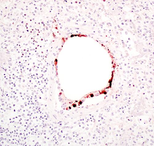
Porcine circovirus-associated disease, porcine respiratory and reproductive syndrome (PRRS), swine influenza (SI), and bacterial bronchopneumonia were diagnosed in a 3-month-old, female pig that died after an 8-day history of fever and neurologic signs. At necropsy, the lungs were non-collapsed, fleshy and had cranioventral consolidation. All lymph nodes were enlarged. Histologically, there was necrotizing bronchointerstitial pneumonia, suppurative bronchopneumonia, lymphoid depletion with inclusion bodies typical of circovirus in lymphoid tissues, and lymphohistiocytic meningoencephalitis. Porcine circovirus-2, PRRS virus, and SI virus were detected (Fig. 1). The latter was typed as H1N2 by whole genome sequencing.
Small Ruminants
Clostridium perfringens type D enterotoxemia and aspiration pneumonia contributed to the death of a 6-year-old Nubian doe. Initially, the doe was slightly bloated and had a frothy mouth. She was treated with sodium bicarbonate, mineral oil, and charcoal. Four hours later she was gasping for breath and died. On necropsy, the rumen pH was 5.5, and there was watery green small intestine and colon content, suggesting rumen acidosis. C. perfringens epsilon toxin was detected in the colon contents, which confirmed a diagnosis of type D enterotoxemia. This doe also had perimortem pulmonary aspiration with charcoal.
Clostridial enterotoxemia type D was diagnosed in a 17-day-old goat that was found dead in the pen. The carcass submitted had brown content in the spiral colon and no other gross or histologic lesions. ELISA for Clostridium perfringens epsilon toxin was positive, which confirmed the diagnosis.
Holiday Schedule
Closed Monday, September 1st for Labor Day.
Employment Opportunities at CAHFS
Davis: Asst/Assoc/Full Professor of Clinical Diagnostic Pathology in the CA Animal Health & Food Safety Laboratory System (CAHFS) and Department of Pathology Microbiology & Immunology. The candidate will function primarily as a diagnostic pathologist in the CAHFS, Davis laboratory. The diagnostic caseload includes a variety of livestock, avian and wildlife species with an emphasis on disease conditions affecting food animal species.
To receive fullest consideration, applications must be received by August 8, 2025; position open until filled.