CAHFS Connection - June 2025

Managing Editor: Kerry Ballinger
Design Editor: Lucy Gomes
Contributors: Anibal Armien, Emma Torii, Francisco Uzal, Javier Asin Ros, Jennine Ochoa, Patricia Blanchard, Raul Resendiz-Pozos, Roberto Olivares, Todd Cornish
Avian
Histomoniasis, or blackhead disease, colibacillosis, and multiple-agent sinusitis and tracheitis were diagnosed in 30-day-old turkey poults from a premises experiencing nasal discharge, swollen heads, and increased mortality. Affected poults had multifocal hepatic necrosis and extensive cecal inflammation and necrosis, both containing protozoal trophozoites consistent with Histomonas meleagridis. The birds also had combinations of sinusitis, tracheitis, and/or airsacculitis and pericarditis. E. coli was isolated from air sac/heart lesions, Bordetella avium was isolated from tracheas, and Pasteurella multocida (cause of fowl cholera) and Trueperella pyogenes were isolated from infraorbital sinuses. The birds also were PCR and serology positive for avian metapneumovirus. This virus contributes to upper respiratory disease and can be immunosuppressive, predisposing to secondary bacterial infections.
Valvular endocarditis was diagnosed in a 10-month-old, female chicken submitted for necropsy. On gross examination, adhered to the mitral valve of the left ventricle of the heart was a large, approximately 0.3 cm3, yellow, friable, round exudate. Aerobic culture of this mass yielded mixed growth of Avibacterium species, Ligilactobacillus salivarius, Escherichia coli and Proteus swarming, hence it was difficult to determine the primary causative agent. Reported bacteria associated with endocarditis in chickens include Staphylococcus aureus, Enterocococus spp., Streptococcus spp., Erysipelothrix rhusiopathiae, and more recently Avibacterium endocarditis.
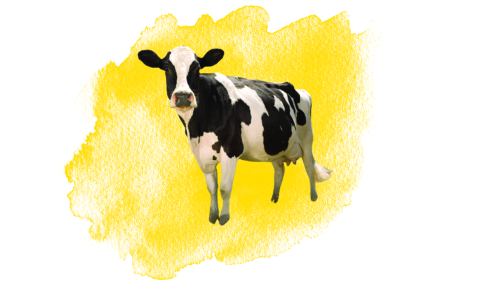
Bovine
A vaccine reaction was the probable cause of death in a 36-day-old female Holstein cow that had signs of anaphylaxis, including head shaking, foamy content in the mouth and nostrils and death within 4 hours after receiving a Salmonella Dublin vaccine. On necropsy, severe pulmonary edema and emphysema, and hemorrhages in multiple organs were observed. Anaphylactic shock typically implies pre-existing exposure to the antigen, which may have originated from a past infection. However, excessive endotoxin in the vaccine, genetic predisposition, or an individual’s exuberant inflammatory response to gram-negative toxins in the vaccine may trigger the reaction.
Blackleg and pneumonia were diagnosed in a 5-month-old beef calf submitted from a herd where 47 beef calves in a group of 350 were down, febrile, and had respiratory signs. Three calves died three days after onset of clinical signs. On gross exam, there was moderate edema and hemorrhage of the brisket, pectoral muscles and caudal esophagus; the lesions had a rancid butter odor. Approximately 70% of the lungs were consolidated and dark red-purple; the visceral pleura was rough and adhered to the thickened, rough pericardium and parietal pleura. Clostridium chauvoei was detected by fluorescent antibody and immunohistochemistry in affected muscle, while Histophilus somni and Mycoplasma bovis were detected in the lung.
Equine
Colonic rupture was diagnosed in a pregnant, 12-year-old, Thoroughbred mare that had a single bout of colic that had resolved two weeks prior to foaling. Immediately after delivering a live foal, the mare began to colic again. Abdominal ultrasound revealed free fluid in the abdomen and distention of the small intestine. Despite supportive care, her condition declined, and she was euthanized. On necropsy, there were approximately 20L of serosanguinous fluid admixed with fecal matter in the abdominal cavity, which was the result of a colonic rupture cranial to the pelvic flexure. The pelvic flexure had an intramural abscess resulting in a markedly narrowed lumen and a 3.5 cm diameter enterolith was lodged in the region. There was also a 3.0 cm diameter subcapsular renal abscess. Streptococcus equi ssp. zooepidemicus was isolated from both abscesses.
Lagomorph
Rabbit hemorrhagic disease virus 2 (RHDV2) infection was the cause of death of a riparian brush rabbit that was found dead. Grossly, the carcass had blood on the face, serosanguineous effusions in the thoracic and abdominal cavities, and pulmonary hemorrhages. Hepatic necrosis was observed histologically. RHVD2 continues being detected periodically in wild and domestic rabbits of Southern and Northern California.
Porcine
Streptococcus suis sepsis caused the death of a 7-day-old piglet that developed a swollen left front leg, trouble breathing, and shaking, less than 12 hours before death. Post-mortem exam and histology revealed suppurative meningitis, arthritis and cellulitis in the left front leg, and bronchointerstitial pneumonia. Streptococcus suis was isolated from meninges, lung and joints.
Small Ruminants
Caseous lymphadenitis and abomasal parasitism were diagnosed in an adult Boer goat found lame and stiff on pasture and euthanized two days after. Postmortem findings included anemia, abundant parasites in the abomasum, and several abscesses measuring ~4 cm in diameter. The abscesses were located on the right side of the head, ventral cervical area, and in the lungs, kidneys, mediastinum, and tracheobronchial lymph nodes. Corynebacterium pseudotuberculosis was isolated from the abscesses.
Clostridium perfringens type D enterotoxemia was the cause of death of a 5-week-old Suffolk lamb that had history of kicking at its belly, running, and falling. The lamb had moderate amounts of yellow fluid in the abdominal and thoracic cavities, and the lungs were mottled. The small intestine had a red mucosa. Histology revealed cerebral perivascular edema and colonic edema. Epsilon toxin was detected in the small intestinal content by ELISA confirming the diagnosis of C. perfringens type D enterotoxemia.
Holiday Schedule
Closed Thursday, June 19th for Juneteenth Holiday
Employment Opportunities at CAHFS
Staff Research Associate I (SRA 1) - San Bernardino - Job #78371
Independently performs sample management, processing, testing, data entry, laboratory maintenance and media preparation for food safety. Performs regulatory testing. The Milk Quality section includes FDA-approved testing such as tests for antibiotic residues, phosphatase, microbial counts, freezing point analysis, sediment determination, somatic cell count, yeast and mold, laboratory pasteurization count, residual bacterial/coliform count and MPN for dairy waters. Participate in the updating and development of analytical procedures, quality control procedures, and Standard Operating Procedures. Responsible for maintaining an inventory, ordering supplies and ensuring the proper operation, calibration and maintenance of laboratory equipment.