CAHFS Connection - October 2025

Managing Editor: Kerry Ballinger
Design Editor: Lucy Gomes
Contributors: Asli Mete, Carmen Jerry, Cassie Powers, Emma Torii, Francisco Uzal, Javier Asin, Karyn Bischoff, Mark Anderson, Nicolas Streitenberger, Patricia Blanchard, Raul Resendiz-Pozos, Tamires Teodoro, Todd Cornish
Avian
Carcinomatosis was diagnosed in an eight-year-old hen with a history of leg paralysis, head bobbing, inappetence, and watery feces. On necropsy, multiple variable size pale nodules were identified in the liver, ovary, and thyroid gland, where neoplastic epithelial cells were observed histologically. Similar neoplastic cells were observed in the small intestinal serosa. Based on these findings carcinomatosis was diagnosed. The most common origin of carcinomatosis in poultry is ovarian carcinoma, although in this case a definitive primary site could not be determined.
Goiter was diagnosed in an adult chicken with wheezing, coughing, and respiratory signs, which progressively worsened over three weeks until the patient was euthanized. On necropsy, the thyroid gland was hyperplastic, and was compressing the trachea, esophagus, and other soft tissues of the neck. Goiter develops, amongst other causes, due to iodine-deficient diets; in this case, the animals were fed broccoli, a well-known goitrogenic agent.
Aquatic
Carp edema virus disease, also known as koi sleepy disease, was the cause of a die-off that affected at least 35 of 52 koi in a domestic pond. The outbreak started shortly after introducing two new koi into the pond. Affected animals had anorexia and erratic behavior prior to death. Two koi carcasses submitted for necropsy had multifocal hemorrhages in the gills (Fig. 1), cornea and vent area. Histologically, both koi had proliferative and necrotizing branchitis; carp edema virus was detected by PCR on tissues of these fish. Carp edema virus is an unclassified poxvirus considered an emerging infectious agent. It can cause high mortality in juvenile and adult koi and common carp.
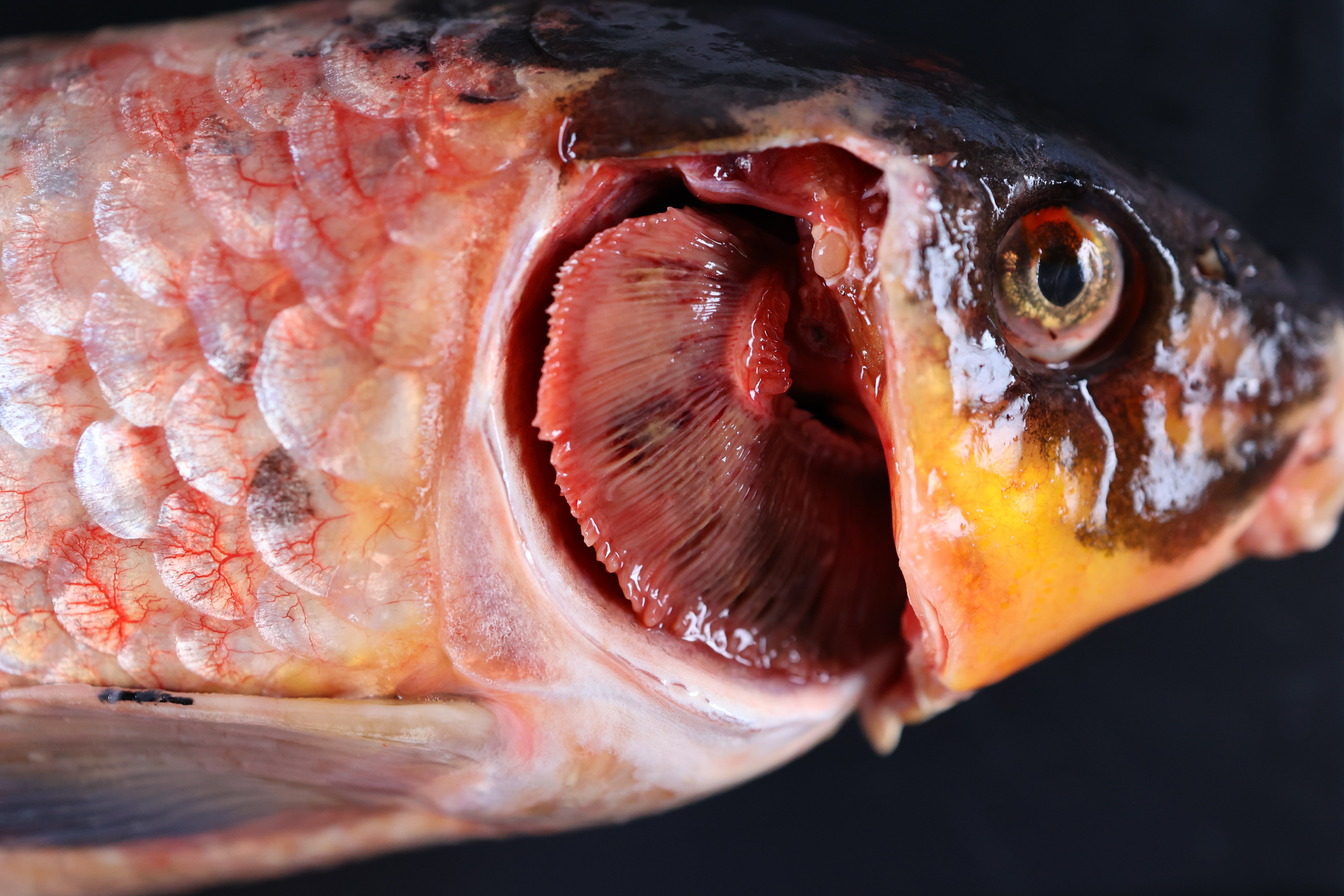
Fig. 1 A koi fish with multifocal hemorrhages in the gills.
Bovine
Sulfur/sulfate toxicosis was the probable cause of illness of an 8-year-old beef cow that was euthanized for doing poorly, and a second beef cow found dead after exhibiting neurologic signs; both were housed on a large pasture. Tissues submitted from both cows did not have gross or microscopic lesions. However, the mineral screen in the eye fluid of both animals showed elevated sulfur levels. Multiple water sources were tested for sulfur/sulfate concentrations; two tested high. Sulfur/sulfate toxicosis can cause variable signs such as diarrhea, poor doing, polioencephalomalacia and death. Drought conditions can exacerbate the occurrence of the intoxication. Ocular fluid can be a good diagnostic specimen for hypomagnesemia, nitrate poisoning, and sodium ion imbalance, and, in this case, it proved helpful to determine sulfur exposure.
Salmonella group D1 septicemia was diagnosed in two, 10-day-old calves from a calf ranch. Both animals had interstitial pneumonia, hepatitis, splenitis and nephritis. Salmonella group D1 (likely S. Dublin) was isolated from multiple organs. Septicemia caused by this organism is more common in 3-12-week-old calves.
Equine
Sand impaction causing colon rupture and peritonitis led to the death of a 10-year-old Quarter Horse mare with a history of colic. Necropsy revealed large amounts of sand, fibrin, and thick tan fluid in the abdominal cavity, and large amount of sand in the right dorsal colon, which had also two perforations. Ancillary testing, including a diarrhea panel, was unremarkable. Sand impaction, often associated with feeding in sandy environments, can result in colic, diarrhea, weight loss and colonic impaction and rupture in horses.
Right dorsal displacement (RDD) of the colon was diagnosed in a Thoroughbred mare with a history of several hours of colic followed by death. On post-mortem examination, the left segments of the large colon were on the right side of the abdomen, and they were twisted, confirming the diagnosis of RDD of the colon. There was also an impaction of the right dorsal colon. RDD is frequently predisposed by impaction of the colon, usually in the pelvic flexure, although in this case the impaction was in the right dorsal colon.
Lagomorph
Intestinal coccidiosis was diagnosed in a 7.5-week-old, male, Californian rabbit (Oryctolagus cuniculus), with a history of lethargy, being off-feed, teeth grinding and diarrhea. It was the fourth rabbit to die in a herd of 45; eight other rabbits were sick with similar signs. Grossly, loose ingesta was noted in the small intestine. Large numbers of coccidia oocysts compatible with Eimeria spp. were detected by fecal floatation (Fig. 2). Intestinal coccidiosis can be particularly pathogenic in young rabbits. However, adults usually have milder clinical signs and are often regarded as resistant. Clinical signs are variable and include diarrhea, sometimes with mucus and blood, dehydration, lethargy, weight loss, stunting, anorexia and death.
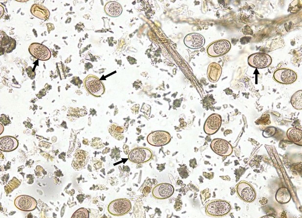
Fig. 2 Fecal flotation showing large numbers of coccidial oocysts compatible with Eimeria spp. (arrows)
Small Ruminants
Copper toxicosis resulted in the death of a female, 2-year-old, Nigerian dwarf goat with a 24-hour history of lethargy before being found dead. On necropsy, there was marked centrilobular to sub-massive hepatic necrosis, and multiple petechial hemorrhages throughout the subcutaneous tissue and heart. The liver copper level was normal, but the kidney copper level was elevated at 20 ppm (normal 3-6 ppm). In some cases, when copper is acutely released from the liver, the liver copper concentration may be within the normal range, but the kidney copper concentration is elevated, such as in this case.
Wildlife
Blunt trauma, anticoagulant rodenticide toxicosis with hemorrhage, and Sarcoptic mange caused the death of an adult male San Joaquin kit fox. The fox was found disoriented and ataxic on a busy road and received emergency treatment, but died in captivity. At necropsy the fox had extensive hair loss and skin crusting over much of the body and tail, hemorrhage in the temporal muscles, a large subdural hematoma, hemothorax and hemoabdomen. Microscopically, skin lesions contained abundant intra-epidermal mites consistent with Sarcoptes sp. Two anticoagulant rodenticides were detected in liver (bromadiolone and difethialone) and these may have exacerbated hemorrhages in head/brain, thorax and abdomen initiated by blunt trauma.
Employment Opportunities at CAHFS
Biotech/Bacteriology Lab Manager (LAB RSCH SUPV 1) – Turlock (81381)
This position is located in Turlock, CA. Supervise and manage diagnostic testing, personnel and related activities in the CAHFS Turlock Biotechnology section and the Bacteriology section related to molecular testing. Responsible for molecular diagnostic testing and laboratory technical support, including processing samples for extraction of DNA and RNA from clinical samples, performance of nucleic-acid based detection, (using real-time PCR, conventional PCR, RT-PCR and amplicon NGS technology) and maintaining documentation records in accordance with standard operating procedures. Supports systemwide Biotechnology efforts under general guidance of systemwide section head to optimize and help author standard operating procedures for PCR-based assays; manage and participate in development and evaluation of new or improved molecular-based detection and diagnostic tools, including RT-PCR and sequencing of isolates; manage the evaluation of new equipment, reagents and supplies; and manage and participate in validation studies to ensure in-house performance of assays. Supervise staff.
Necropsy Technician (SRA 2 NEX) - Davis (81353)
Under minimal supervision, handle animals, perform necropsies, process fixed tissue obtained from animal necropsy procedures, and trim formalin fixed tissues with adherence to established protocols. Provide high level technical assistance, data entry, and laboratory maintenance for necropsy facilities.
Poultry Medicine and Diagnostics Residency Program - Turlock, CA
The California Animal Health & Food Safety Laboratory System (CAHFS), School of Veterinary Medicine, University of California, Davis is offering a two-year Poultry Medicine and Diagnostics Residency Program at its Turlock Laboratory. This program is designed to provide training in poultry disease diagnostics and prevention, production medicine with special emphasis on flock health and commercial poultry and communication with industry. The program is recognized as an approved training program by the American College of Poultry Veterinarians. The training involves extensive poultry diagnostic casework. Casework is supplemented by weekly case conferences, seminars, lectures, and rotations through specialty laboratories including bacteriology, immunology, histopathology, and virology.