CAHFS Connection - January 2025

Managing Editor: Kerry Ballinger
Design Editor: Lucy Gomes
Contributors: Emma Torii, Francisco Uzal, Javier Asin, Jennine Ochoa, Mark Anderson, Wendi Jackson
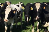
The California Animal Health and Food Safety (CAHFS), in collaboration with state and federal government partners is working around the clock to support the state-wide effort to control highly pathogenic H5N1 (bird flu) influenza virus in poultry and cattle. The main goals of this effort are to protect animal and human health and food safety, by providing high quality diagnostic testing for livestock and poultry.
This is an outbreak of unprecedented scale that has already affected at least 650 dairies, and more than 25 commercial and 6 backyard poultry flocks in California. CAHFS is putting resources available from our 4 branches (Davis, Turlock, Tulare and San Bernardino) towards this extraordinary effort. We are grateful to CAHFS staff for their incredible commitment and professionalism, and are extremely fortunate to have skilled and dedicated employees.
We are also fortunate to be a member of the National Animal Health Laboratory Network (NAHLN), a consortium of veterinary diagnostic laboratories authorized to perform regulatory testing at the same standards as the USA’s national reference laboratory. The NAHLN exists in part to provide surge capacity for outbreaks, and is providing direct and indirect assistance to CAHFS and to California so that collectively, the testing needs for this outbreak can be met.
Consumers are reminded by the FDA that pasteurization is 100% effective in destroying highly pathogenic H5N1 influenza virus in milk. The Milk Quality (MQ) section of CAHFS is the designated Central Laboratory for California in the Food and Drug Administration Pasteurized Milk Ordinance program and plays a major role in monitoring the safety of dairy products. The MQ section helps ensure that California's milk, milk products, and products resembling milk are safe, wholesome and meet microbial, pasteurization, and other standards.
Dairy and poultry owners are encouraged to protect their herds and flocks by increasing their biosecurity practices. Recommended biosecurity practices can be found on the CDFA and USDA websites.
Dr. Ashley Hill, DVM MPVM PhD
CAHFS Director
Submission of milk and serum samples for H5N1 influenza testing. Please note that CAHFS cannot accept milk or serum samples for H5N1 influenza testing (molecular or serology testing) if the submission form accompanying the samples does not include a National Premises ID Number (NPIN). If the location does not already have a NPIN, the submitter can call the California Department of Food and Agriculture Premises ID (866-325-5681) to obtain one. A submission reason must also be clearly indicated on the form, such as Herd Monitoring (for general disease surveillance), Interstate Movement, etc.
New!
CAHFS-Davis is able to test bovine milk and serum for antibodies against Influenza A virus by ELISA. Only milk that is Avian Influenza PCR negative can be tested by ELISA. Consult the shipping details for this testing under Tests & Fees on the CAHFS website prior to sending in specimens or call the lab for details.
Avian
Avian pulmonary proteinosis was diagnosed in a 17-month-old chicken with a two-day history of lethargy and anorexia prior to death. The lungs were wet and mottled pale to dark pink on gross examination. On histology, there were large deposits of material in air spaces, consistent with surfactant. Avian pulmonary proteinosis has been sporadically reported in birds, including chickens, and is caused by abnormal accumulation of surfactant in the lungs. In some cases, this is an incidental finding, but in severe cases such as this one, it can lead to respiratory failure and death.
Pseudomonas aeruginosa septicemia was diagnosed in eight, 16-day-old quail chicks presented with a history of poor viability after hatching, bloody noses, sneezing, and lethargy in the last two hatches.

On necropsy, the livers and spleens were enlarged and mottled red to brown. Microscopically, there was splenic and hepatic necrosis. These changes are compatible with septicemia. P. aeruginosa was isolated from the livers in pure culture. This microorganism grows well in warm, humid climates and in this case, it is believed that the incubators were contaminated.
Bovine
H5N1 Avian influenza (AI) virus has been recently diagnosed as the cause of a marked uptick in abortions and stillbirths on several dairies. On necropsy, fetuses had pale-streaked hearts, dark red lungs with prominent interlobular septa, and enlarged livers. Microscopic lesions included myocardial necrosis, mineralization, and lymphocytic inflammation. The lungs and livers had vasculitis. A few fetuses had lymphocytic meningoencephalitis. H5N1 AI virus was detected by PCR on lung swabs. Affected tissues were positive for Influenza A on immunohistochemistry.
Leptospira interrogans serovar pomona was the cause of stillbirth in a crossbred beef calf from a herd of 100 pastured cattle where four stillbirths and three deaths occurred shortly after birth. Field necropsy revealed marked, diffuse icterus, swollen kidneys, pale liver, and enlarged spleen.

Histology revealed mild suppurative nephritis. The L. pomona titer in the dam was >1:3200 while the other 4 serovars were negative at 1:100. The herd had not been vaccinated against leptospirosis. PCR, immunohistochemistry, and silver stains failed to detect the organism; Leptospira organisms can be multifocal in tissues and missed on sampling, which supports the value of serology on dam sera on abortion submissions.
Equine
Epiploic foramen entrapment and strangulation of the large colon were diagnosed in a five-year-old Thoroughbred mare with a 24-hour history of colic that was euthanized after failing to respond to medical treatment. At necropsy, approximately three meters of small intestine, including the distal jejunum and proximal ileum, were incarcerated (from left to right) through the epiploic foramen. The incarcerated segment of intestine was very congested, hemorrhagic and edematous. In addition, the anterior part of the jejunum was wrapped around the sternal and diaphragmatic flexures of the large colon producing a constriction and obstruction of this organ.
Actinobacillus equuli ssp. equuli neonatal septicemia was the cause of death of a one-day-old, male, miniature donkey that was lethargic since birth. At necropsy, the lungs were non collapsed and histologically there was multifocal hemorrhage and necrosis with intralesional bacteria in many organs. A. equuli ssp. equuli was isolated from liver, lung, and spleen, and there were no IgG in serum, which is consistent with failure of passive transfer. A. equuli ssp. equuli is part of the normal microbiota of the gastrointestinal and reproductive systems. Fetal/neonatal infection can occur in uterus, during parturition or through the umbilicus or small wounds. Poor colostrum intake/failure of passive transfer is the main predisposing factor.
Pigs
Colonic sand impaction and mesocolon twist resulted in the death of an eight-month-old Duroc barrow. The hog was wheezing, teeth grinding, and vomiting in the 36 hours prior to death. Necropsy revealed a 360o twist of the mesocolon and the entire spiral colon was bright to dark red, gas distended with fibrin over the serosa, and had dark red-brown content. Distal to the twist, a 32 cm segment of colon was impacted with dry gray sand and there was focal pressure necrosis of the wall. The twist was most likely secondary to the sand impaction.
Valvular endocarditis of the right atrioventricular valve was diagnosed in a three-month-old Yorkshire pig that died after a four-day period of lethargy, anorexia and pale mucous membranes. Fresh heart was submitted for examination and testing, and multifocal to coalescing, gritty-to-soft, cauliflower-like yellow-to-green masses were observed in the right atrioventricular valve. Dense fibrinosuppurative inflammation admixed with large colonies of cocci were observed histologically, and Streptococcus suis and Escherichia coli were isolated.
Small Ruminant
Acute bacterial bronchopneumonia was the cause of death of a juvenile Nubian goat that was found in the morning without observed prior signs of illness. Grossly, there was pulmonary cranioventral consolidation with fibrin over the pleura. Histologically, there was severe acute suppurative inflammation with “oat cells” and bacterial colonies. Streptococcus sp., Klebsiella aerogenes, and Escherichia coli were recovered from the lung. No Mycoplasma spp. was isolated. The presence of “oat cells” suggests that other bacteria, in particular members of the family Pasteurellaceae, such as Mannheimia haemolytica, may have played a role. In this case, the poor postmortem condition of the carcass could have hindered the bacteriology results.
Wildlife
Rabies was diagnosed in a grey fox that was found dead. On necropsy, this animal had punctures of the skin and underlying musculature in the thorax, fractured ribs, hemothorax, and pulmonary hemorrhage. These changes were considered to be compatible with predation. Microscopically, there was lymphoplasmacytic encephalitis with intracytoplasmic inclusion bodies characteristic of rabies. Rabies was confirmed by immunofluorescence.
Tularemia was diagnosed in an immature female California ground squirrel which was exhibiting severe distress before being euthanized. Necropsy and histologic examination revealed disseminated necrotic foci in lung, liver, spleen and lymph nodes, and multiple hydatid cysts in the liver. Francisella tularensis was detected by PCR on tissue samples. Immunohistochemistry for this microorganism was also positive. The hydatid cysts in liver were considered incidental.
Holiday Schedule
- New Year's Holiday, Wednesday, January 1, 2025 - Closed
- Martin Luther King Day, Monday, January 20, 2025 - Closed
CAHFS is Hiring!
Davis
Sample Processing Technician (LAB AST 3) - Job# 75147
Perform specimen processing for chemical analysis of regulated substances in biological substances. Prepare various reagents and solutions. Perform sample extraction and testing methods in accordance with Standard Operating Procedures. Maintain laboratory inventory of supplies, samples, and materials.
