


CAHFS Connection - January 2026

Managing Editor: Kerry Ballinger
Design Editor: Lucy Gomes
Contributors: Carmen Jerry, Cassie Powers, Emma Torii, Francisco Uzal, Javier Asin Ros, Karyn Bischoff, Melissa Macias Rioseco, Patricia Blanchard, Simone Stoute
Avian
Salmonella Enteritidis (SE) was isolated from 28-day-old commercial broilers with a history of stunting. On necropsy, pericarditis, perihepatitis, synovitis, osteomyelitis, and airsacculitis were identified. SE was isolated in pure culture from the heart, liver, and bone, and E. coli was isolated from the upper respiratory tract and joints. SE is of significant public health importance, as it is a well-recognized foodborne pathogen. In poultry, SE can colonize the intestinal and reproductive tracts, enabling vertical and horizontal transmission within flocks. Clinical disease in young birds may result in septicemia and fibrinous inflammation, leading to increased mortality, particularly when compounded by concurrent bacterial infections or environmental stressors. Subclinical infections are also common and can contribute to environmental contamination and ongoing transmission. This case underscores that, beyond its zoonotic implications, SE can cause septicemia with notable morbidity and mortality in poultry.
Acute fowl cholera was diagnosed in a flock of 25-week-old Ring-necked pheasants (Phasianus colchicus) with a history of drooping heads, crouching down and dying suddenly. The mortality was approximately ten birds per week, and the morbidity was 0.5% in a flock of 20,000 birds. On necropsy, mild airsacculitis, enlarged mottled spleen, reddened proventricular mucosa and scant mucoid exudate in the infraorbital sinuses was observed. Pasteurella multocida, the causative agent of fowl cholera, was isolated from the liver, spleen, brain, and lung. Necrosis of liver, spleen and cranial bones was visible microscopically. Clinical signs of fowl cholera vary with species, age, and the immune status of the birds. In acute fowl cholera, sudden onset of high mortality is often the first sign. In chronic fowl cholera, lesions are localized to wattles, joints, cranial bones, and middle ear among others.
Intestinal blockage secondary to severe tapeworm infection was diagnosed in a 1-year-old Swedish Flower rooster that died following a five-day illness, including green stools. On postmortem examination, the chicken was thin and had a prominent keel bone. Within the distal jejunum, ileum, and both ceca were numerous tapeworms that blocked the lumen of those organs. Additionally, fecal flotation identified a high load of coccidia oocysts, Heterakis/Ascaridia eggs, and tapeworm eggs. Most cases of severe tapeworm infections cause weight loss, poor growth, decreased egg production, and lethargy, as the worms attach to the intestinal wall and absorb nutrients. A severe infestation can also cause intestinal blockage and death, as seen in this case.
Bovine
Choke and salmonellosis were diagnosed in a 4-day-old Angus bull calf with history of weakness. The most significant postmortem findings were fibrinous peritonitis and esophageal obstruction by a white tablet located within the distal third of the esophagus (Fig. 1). The associated esophageal mucosa was ulcerated. Esophageal choke in calves is often caused by foreign objects, feed, or, in young calves, congenital issues such as megaesophagus. Signs include anxiety, extended neck, excessive salivation, and regurgitation of milk. Additionally, there was acute, severe segmental enteritis caused by Salmonella Typhimurium (Fig. 2). This microorganism is a common cause of diarrhea in calves < 2-months-old.

Figure 1. A white chalky tablet located in the distal 1/3rd of the esophagus was causing ulceration (arrow) and obstructing the organ.

Figure 2. The intestinal mucosa was necrotic and covered by a fibrino-necrotizing pseudomembrane.
Mycoplasma bovis infection within the atlanto-occipital joint was diagnosed in an 11-month-old heifer with a history of inability to stand following treatment. On necropsy, the left side of the neck at the level of the atlanto-occipital joint had a poorly demarcated abscess that extended into the musculature, lymph nodes, and spinal canal. M. bovis was detected by PCR in neck tissue and joint fluid. Septic arthritis of the atlanto-occipital joint in cows is primarily caused by bacterial infection, which can occur through hematogenous spread, direct trauma, or extension of a nearby infection. Common bacterial culprits include Trueperella pyogenes, Staphylococcus spp., Streptococcus spp., Escherichia coli, and M. bovis.
Camelid
Disseminated coccidioidomycosis (Valley fever) was the cause of death of an 11-year-old female alpaca. Grossly, the carcass was severely emaciated and there were multifocal to coalescing, caseous and gritty, tan nodules in the skin around the right ear, multiple subcutaneous and visceral lymph nodes, lungs (Fig. 3), heart, liver, spleen, and omentum. Histologically, these nodules corresponded to pyogranulomatous inflammation with necrosis and numerous fungal spherules with endospores consistent with Coccidioides spp. (Fig. 4).
Valley fever is endemic in California and is a zoonotic disease. It may be caused by two different species of Coccidioides: C. immitis and C. posadasii. The fungus is present in dusty, sandy soils, and is most frequently acquired through inhalation; from the lungs, it can disseminate systemically though the blood. Coccidioidomycosis is relatively frequent in alpacas, llamas and other domestic camelids in the southwestern United States, and pneumonia is a very usual outcome.
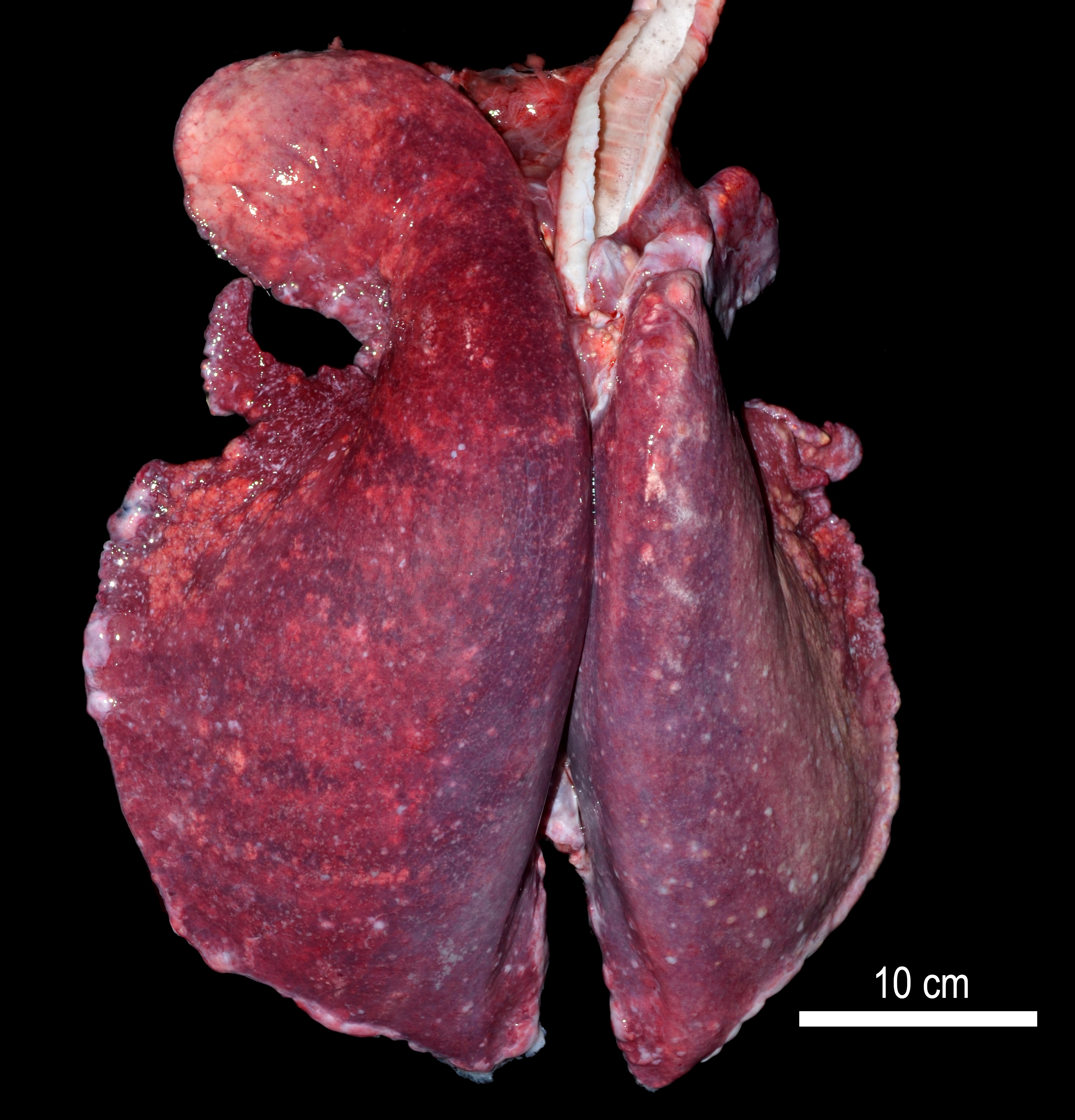
Figure 3. Lungs from alpaca with multifocal pyogranulomas due to Valley Fever.

Figure 4. Spherules in the lung of an alpaca with valley fever.
Equine
Lymphoma was diagnosed in an 18-year-old mare with a 6-month history of hindlimb incoordination, weakness, falling over and weight loss. On necropsy, there was lymphoma infiltrating the brain, spinal cord, peripheral nerves, aorta, pulmonary artery, stomach, heart and a lymph node. Central nervous system involvement of equine lymphoma is rare, although it can be a manifestation of multicentric lymphoma. Additionally, there were bilateral skin ulcerations of the hock, which likely occurred from the horse falling over. In the left hindlimb was marked cellulitis and tenosynovitis, calcaneal bursa bursitis, and septic arthritis of the fetlock, which likely was secondary to infection from the ulcerated skin.
Cirrhosis due to pyrrolizidine alkaloid (PA) toxicosis was diagnosed in an adult gelding. On physical examination, the horse was markedly thin with poor hoof conformation and dentition. On necropsy the liver was shrunken and had multifocal, variably sized firm nodules that were continuous with the hepatic parenchyma on cut section. On histology, the hepatic parenchyma had variably sized and well demarcated nodules outlined by marked periportal bridging fibrosis and occasional megalocytosis. Based on these findings, a presumptive diagnosis of PA toxicosis was established. PA toxicosis is a chronic disease that often results in hepatic failure. It is caused by many toxic plants, including the genus Senecio, Crotalaria, Heliotropium, Amsinckia, Echium, Cynoglossum, and Trichodesma. Clinical signs include loss of condition, anorexia, dullness, and constipation or diarrhea.
Small Ruminants
Epizootic hemorrhagic disease virus (EHDV) infection was diagnosed in a wild mule deer from an area experiencing increased mortality and sick appearing deer. Gross exam revealed pulmonary hemorrhages and edema, and fibrin over abdominal serosas. Histopathologic findings included multifocal vasculitis affecting the lung and brain, pulmonary edema with hemorrhage, and fibrinous peritonitis and perihepatitis. In addition, the lungs had mild granulomatous pneumonia associated with intralesional nematode parasites. Ectoparasite infestation with Lipoptena sp. (deer ked) and Dermacentor sp. (tick) was also noted. EHDV was detected by PCR. This virus is transmitted by biting midges (Culicoides spp.) and can cause acute and severe disease in wild and domestic ruminants, characterized by fever, edema, hemorrhages, and sudden death.
Wildlife
Bromethalin toxicosis was diagnosed in a litter of 5, 1-month old pre-weaned feral kittens presented to a rescue group. Kittens developed progressive neurologic dysfunction over three days and one died. Living kittens were obtunded and tetraparetic, with some voluntary movement observed in all four limbs. Serum desmethylbromethalin concentrations for the 4 living kittens ranged from 160-290ppb. Desmethylbromethalin is the active metabolite of bromethalin, a common and neurotoxic rodenticide product. One kitten began to improve with treatment over the next several days and was soon able to stand and eat on her own. Within a week, she became ambulatory and she continued to improve. Her serum desmethylbromethalin concentration dropped from initial 160ppb to 8.8 ppb by 18 days after submission and 2.47 ppm 9-days later.
Holiday Schedule
- New Year’s Holiday, Thursday January 1, 2026 – Closed
- Martin Luther King Day, Monday January 19, 2026 – Closed