


CAHFS Connection - June 2026

Managing Editor: Kerry Ballinger
Design Editor: Lucy Gomes
Contributors Cassie Powers • Emma Torii • Francisco Uzal • Javier Asin Ros • Patricia Blanchard • Todd Cornish
Avian
Articular and visceral gout in a Welsummer hen
Articular and visceral gout were diagnosed in a 4-year-old Welsummer hen with a history of lameness, joint swelling, and decreased activity. Both hindlimbs were diffusely thickened with small to moderate amounts of white, dry, caseous material in the joints. Both kidneys were covered by numerous <2 mm in diameter tan foci. On histopathology, urate tophi were observed in joints and kidneys. Several predisposing factors have been associated with gout, including severe renal disease, dehydration, postrenal obstruction, excessive dietary protein, low calcium diets, vitamin A deficiency, inherited enzyme defects, exposure to mycotoxins, and nephrogenic infectious bronchitis virus.
Poorly differentiated neoplasm with mycotic proventriculitis in a green-cheek conure
A poorly differentiated tumor and mycotic proventriculitis was diagnosed in an adult female green-cheek parakeet (conure) with a history of sudden death.
Gross evaluation showed a severely enlarged proventriculus with a large blood clot in the lumen. On microscopic evaluation, the wall of the proventriculus was infiltrated by a large poorly differentiated neoplasm. On the surface of the neoplasm, was a focus of infection with fungal hyphae, which on GMS special stain (Fig. 1), is most suggestive for Candida spp. organisms.

Bovine
Systemic Mycoplasma bovis infection in beef/dairy cross calves
Two 25-day-old beef/dairy cross calves were diagnosed with systemic Mycoplasma bovis infection. At necropsy, both calves had bronchopneumonia and polyarthritis, affecting carpal, fetlock, stifle and hock joints. One calf also had bilateral otitis media. M. bovis was isolated and detected by PCR from lungs, joints and middle ears. At the home calf ranch, more than 15 calves were similarly affected, most with significant lameness due to the polyarthritis. Co-infection with Trueperella pyogenes was present in some joints.
Navel Ill with polyarthritis in a bull calf
Navel-ill and polyarthritis was diagnosed in a 1-month-old bull calf. Gross evaluation showed the umbilical and portal veins were firm and contained abundant pus extending to the liver (Fig. 2). There were multiple abscesses throughout the right and medial lobes of the liver. All evaluated joints contained abundant fibrin and fluid (Fig. 3).
Hemolytic Escherichia coli, Streptococcus species, and Trueperella pyogenes were isolated from the umbilicus and joints. Navel ill is a common infection of the umbilical stump in newborn calves resulting from bacteria entering the body through the navel. It often leads to abscesses and infection of the joints (polyarthritis). It typically occurs within the first one to two weeks of life; however, older calves can still be affected due to delayed healing and/or slow drying of the umbilicus.

Equine
Demodicosis in a Paint horse
Demodicosis was diagnosed in an eyelid biopsy from a 17-yr-old Paint Horse. The horse had a history of a small, ulcerated lesion on the eyelid that was treated with cryotherapy and local 5-fluorouracil, which had caused an inflammatory reaction. On histology, there was dilation of the ducts of the Meibomian glands, with multiple Demodex mites. In this case, Demodex caballi was suspected given the location of the mites.
Cervical fracture in a Dutch warmblood mare
A Dutch Warmblood mare sustained a cervical fracture at a show jumping event. The horse refused to jump, unseating the rider, then panicked and ran into the road where it skidded headfirst into a passing car. On necropsy, there was a complete, closed, heavily comminuted fracture of the fourth cervical vertebra (Fig. 4), involving both the body and the laminae, with severe impingement of the cervical spinal cord. Severe hemothorax was also noted.

Small ruminants
Mammary gland squamous cell carcinoma in a Nigerian dwarf goat
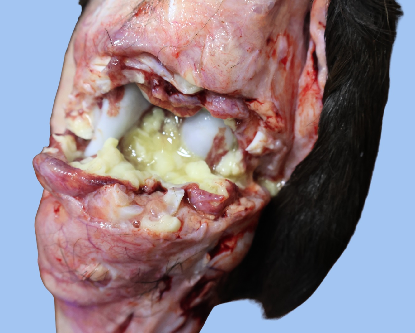
Mammary gland squamous cell carcinoma was diagnosed in a seven-year-old Nigerian dwarf doe goat. On gross examination, the right mammary gland was markedly enlarged, with a large, necrotic, malodorous center that contained a large amount of yellow-brown, viscous exudate, surrounded by a thick white capsule. On histology, the mass was diagnosed as a squamous cell carcinoma, with a large, infected, necrotic center. Squamous cell carcinomas are reported as the most common epithelial tumor in goats, with udder and perianal skin being the most common sites. In this case, there was no obvious skin involvement, hence it was possible the squamous cell carcinoma arose from the mammary gland itself.
Degenerative joint disease and uterine adenocarcinoma in a Nigerian Dwarf goat

Degenerative joint disease (DJD) and uterine adenocarcinoma were diagnosed in a 13-year-old female Nigerian dwarf goat with history of rocking on her front legs due to pain.
On necropsy, both elbow joints had large amounts of straw-colored fluid, and numerous cartilage and bone ulcers. Based on these findings, negative bacteriology result, and lack of additional microscopic lesions, a diagnosis of DJD was made. DJD is relatively common in older animals and elbow OA seems to be particularly prevalent.
Additionally, on gross evaluation the uterus had multiple firm tan nodules (Fig. 5), which on histology were diagnosed as adenocarcinoma nodules. This neoplasia of the uterus is rarely diagnosed in ruminants, but it is usually highly metastatic and locally invasive.
Other species
Hepatic sarcoma with cestodiasis and Filobacterium rodentium pneumonia in a rat
Cestodiasis with associated hepatic and metastatic sarcoma, and Filobacterium rodentium (formerly known as CAR bacillus) pneumonia was diagnosed in a two-year-old male intact rat with history of respiratory distress and sudden death. On necropsy, the left lateral liver lobe was replaced by a multinodular neoplasm in the center of which were several tapeworms (Fig. 6A). Additional masses were present throughout the mesentery and within the abdominal wall. Histopathology of these masses confirmed a sarcoma. While many tapeworm infections are asymptomatic, heavy infections can cause an enlarged liver and lead to tumor development.
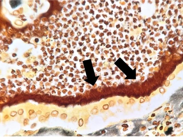
Microscopic evaluation of the lungs revealed bronchopneumonia with bronchiectasis and filamentous bacteria parallel to the bronchiolar cilia, consistent with Filobacterium rodentium (formerly CAR Bacillus) (Fig. 6B).

Holiday Schedule
Juneteenth Holiday: Friday June 19, 2026 - Closed
Employment Opportunities
None listed at this time.